Contents

SynopsisCodesLook ForDiagnostic PearlsDifferential Diagnosis & PitfallsBest TestsManagement PearlsTherapyDrug Reaction DataReferences

View all Images (10)

Pancreatic carcinoma
Other Resources UpToDate PubMed

Pancreatic carcinoma

Contributors: Peter Krasniak BS, Jennifer J. Findeis-Hosey MD
Other Resources UpToDate PubMed

Synopsis

Pancreatic carcinoma typically arises from the exocrine pancreas and is the fourth leading cause of cancer death in men and women in the United States. Ductal adenocarcinoma and its subtypes represent 85% of all pancreatic cancers, with the remainder due to mucinous cystic neoplasms, pancreatoblastoma, serous cystadenoma, neuroendocrine tumors, and intraductal papillary-mucinous neoplasms. Pancreatic cancer is more common in men than women, and incidence increases with advancing age.

Risk factors for development of pancreatic cancer include a first-degree relative with exocrine pancreatic cancer, hereditary pancreatitis, nonhereditary chronic pancreatitis, and cigarette use.

Patients typically present with advanced disease, and signs and symptoms include pain, jaundice, and weight loss.

Codes

ICD10CM:
C25.9 – Malignant neoplasm of pancreas, unspecified

SNOMEDCT:
372142002 – Carcinoma of pancreas

Look For

Subscription Required

Diagnostic Pearls

Subscription Required

Differential Diagnosis & Pitfalls

To perform a comparison, select diagnoses from the classic differential

Subscription Required

Best Tests

Subscription Required

Management Pearls

Subscription Required

Therapy

Subscription Required

Drug Reaction Data

Subscription Required

References

Subscription Required

Last Reviewed:12/12/2017
Last Updated:07/06/2022
Copyright © 2024 VisualDx®. All rights reserved.
Pancreatic carcinoma
A medical illustration showing key findings of Pancreatic carcinoma () : Abdominal pain, Diarrhea, Hepatomegaly, Jaundice, Nausea, Bilirubin elevated, Malaise, Trypsin elevated, Anorexia, Pruritus
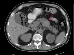
Imaging Studies image of Pancreatic carcinoma - imageId=6846876. Click to open in gallery.  caption: 'Axial non-contrast CT image demonstrates a relatively hyperattenuating mass within the body of the pancreas. Pathology was consistent with pancreatic neuroendocrine tumor.'
Axial non-contrast CT image demonstrates a relatively hyperattenuating mass within the body of the pancreas. Pathology was consistent with pancreatic neuroendocrine tumor.
Copyright © 2024 VisualDx®. All rights reserved.