Contents

SynopsisCodesLook ForDiagnostic PearlsDifferential Diagnosis & PitfallsBest TestsManagement PearlsTherapyReferences

Information for Patients

View all Images (87)

Atopic dermatitis in Infant/Neonate
See also in: Cellulitis DDx,External and Internal Eye
Other Resources UpToDate PubMed

Atopic dermatitis in Infant/Neonate

See also in: Cellulitis DDx,External and Internal Eye
Contributors: Azeen Sadeghian MD, Sophia Delano MD, Susan Burgin MD
Other Resources UpToDate PubMed

Synopsis

Atopic dermatitis (eczema) is a chronic, relapsing, pruritic condition characterized by (1) pruritus (itch); (2) facial and extensor involvement during infancy that changes to flexural involvement in late childhood; (3) a chronic relapsing course; and (4) a personal or family history of atopy (atopic dermatitis, food allergies, allergic rhinitis, and/or asthma).

Infants and children are most often affected, with 85% of cases appearing in the first year of life, and 95% of cases appearing by 5 years. Uncommonly, the condition may persist into, or even arise in, adulthood. Less than 1% of adults are affected by atopic dermatitis.

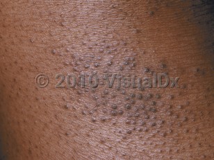
In infants, the disease involves primarily the face, scalp, torso, and extensor aspects of extremities. In children and adults, the disease usually involves chiefly the flexural aspects of extremities, but it may be more generalized. In adults, flexural skin may be clear, and disease may be focal or widespread. Follicular patterns of atopic dermatitis (ie, follicular eczema) are more common in persons with darker skin colors.

Atopic dermatitis may be categorized as follows:
  • Acute – erythema, vesicles, bullae, weeping, crusting
  • Subacute – scaly plaques, papules, round erosions, crusts
  • Chronic eczema – lichenification, scaling, hyper- and hypopigmentation
The cause of atopic dermatitis is unknown. Genetic and environmental predisposing factors exist. Multiple loci have been associated with atopic dermatitis. A family history of atopic dermatitis is common.

Intense pruritus is a hallmark of atopic dermatitis. Scratching leads to lichenification (skin thickening). Impaired barrier function results in increased transepidermal water loss and susceptibility to secondary bacterial and viral infections. Patients with atopic dermatitis are prone to impetiginization with Staphylococcus aureus. Secondary infections with herpes simplex virus (eczema herpeticum), Coxsackie viruses (eczema coxsackium), vaccinia virus (eczema vaccinatum), or molluscum contagiosum may occur.

Patients with atopic dermatitis have difficulties in retaining skin moisture and suffer from xerosis (dry skin). Environmental triggers, such as heat, humidity, detergents / soaps, abrasive clothing, chemicals, smoke, and even stress, tend to aggravate the condition. Latex allergy and nickel allergy occur more often in persons with atopic dermatitis. Additionally, patients with atopic dermatitis have been found to be more likely to have positive patch test results to products commonly found in topical treatments, including cocamidopropyl betaine, wool alcohol / lanolin, and tixocortol pivalate. Allergy to eggs, cow's milk, or peanuts is common. There may be a relationship between atopic dermatitis and the development of aspirin-related respiratory disease.

Atopic dermatitis is increasing in the developed world. In the United States, about 10% of children may be affected by atopic dermatitis, but the majority of these cases are mild.

Codes

ICD10CM:
L20.9 – Atopic dermatitis, unspecified

SNOMEDCT:
24079001 – Atopic dermatitis

Look For

Subscription Required

Diagnostic Pearls

Subscription Required

Differential Diagnosis & Pitfalls

To perform a comparison, select diagnoses from the classic differential

Subscription Required

Best Tests

Subscription Required

Management Pearls

Subscription Required

Therapy

Subscription Required

References

Subscription Required

Last Reviewed:03/22/2017
Last Updated:11/21/2023
Copyright © 2024 VisualDx®. All rights reserved.
Patient Information for Atopic dermatitis in Infant/Neonate
Print E-Mail Images (87)
Contributors: Medical staff writer
Premium Feature
VisualDx Patient Handouts
Available in the Elite package
  • Improve treatment compliance
  • Reduce after-hours questions
  • Increase patient engagement and satisfaction
  • Written in clear, easy-to-understand language. No confusing jargon.
  • Available in English and Spanish
  • Print out or email directly to your patient
Copyright © 2024 VisualDx®. All rights reserved.
Atopic dermatitis in Infant/Neonate
See also in: Cellulitis DDx,External and Internal Eye
A medical illustration showing key findings of Atopic dermatitis : Bilateral distribution, Dry skin, Erythema, Excoriated skin lesion, Flexural distribution, Lichenified plaque, Ocular pruritus, Pruritus
Clinical image of Atopic dermatitis - imageId=214627. Click to open in gallery.  caption: 'A close-up of follicular papules with scale.'
A close-up of follicular papules with scale.
Copyright © 2024 VisualDx®. All rights reserved.