Calcified nodules of the heel represent dystrophic calcification of the skin (calcinosis cutis) following trauma, mostly due to needle sticks during the neonatal period. Lesions occur in both normal infants and in high-risk neonates with a history of repeated heel sticks. It is believed alkaline phosphatase is released during the needle stick, leading to an altered local pH, with precipitation of calcium salts. Serum calcium and phosphate levels are normal.
Calcified nodules begin as small yellow-to-white nodules that slowly increase in size and migrate through the epidermis to the skin surface. Lesions typically appear between 4 and 12 months of age and often resolve spontaneously between 18 and 30 months. Most lesions are asymptomatic, but some may cause local irritation or pain.
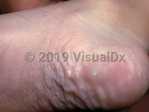
Calcified nodules of the heels
Alerts and Notices
Important News & Links
Synopsis

Codes
ICD10CM:
L94.2 – Calcinosis cutis
SNOMEDCT:
255003 – Calcified nodule
L94.2 – Calcinosis cutis
SNOMEDCT:
255003 – Calcified nodule
Look For
Subscription Required
Diagnostic Pearls
Subscription Required
Differential Diagnosis & Pitfalls

To perform a comparison, select diagnoses from the classic differential
Subscription Required
Best Tests
Subscription Required
Management Pearls
Subscription Required
Therapy
Subscription Required
References
Subscription Required
Last Updated:01/24/2022
Calcified nodules of the heels