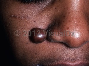
Chondroid syringoma
Alerts and Notices
Important News & Links
Synopsis

Chondroid syringoma, or mixed tumor of the skin, is a rare benign tumor of the sweat glands. The term mixed tumor refers to the presence of epithelial and mesenchymal elements derived from either eccrine or apocrine glandular tissue.
Chondroid syringomas present as small (0.5-3 cm), painless, slow-growing masses in the head and neck region. They are typically solitary dermal or subcutaneous nodules found in middle-aged men, although cases of multiple tumors, larger tumors, and tumors in various locations, across ages, and in both sexes have been reported.
Malignant transformation of mixed tumors of the skin is very rare. Malignant forms tend to grow rapidly and occur more commonly on the lower extremities of younger females. Metastasis to regional lymph nodes, bone, and visceral organs may occur.
Chondroid syringomas are rare, representing less than 0.1% of cutaneous tumors. Given their low rate of incidence and nonspecific clinical presentation, they are rarely a preoperative diagnosis of skin tumors. Definitive diagnosis requires total excision and histopathologic examination.
Codes
ICD10CM:
D23.9 – Other benign neoplasm of skin, unspecified
SNOMEDCT:
302828001 – Syringoma of skin
D23.9 – Other benign neoplasm of skin, unspecified
SNOMEDCT:
302828001 – Syringoma of skin
Look For
Subscription Required
Diagnostic Pearls
Subscription Required
Differential Diagnosis & Pitfalls

To perform a comparison, select diagnoses from the classic differential
Subscription Required
Best Tests
Subscription Required
Management Pearls
Subscription Required
Therapy
Subscription Required
References
Subscription Required
Last Updated:01/07/2019

