- Angioleiomyoma – This most common type of CL originates from the smooth muscle of vessel walls.
- Piloleiomyoma – The next most common type of CL evolves from the arrector pili muscle associated with hair follicles.
- Genital leiomyoma – The least common type of CL is derived from smooth muscle associated with the external genitalia and nipple-areolar complex.
Cutaneous angioleiomyoma typically presents as a single, slowly enlarging papule or nodule on the leg of a middle-aged woman. However, men are also affected, and pediatric cases have been reported. Angioleiomyomas have been reported on the trunk, face, and upper extremities. While lower extremity lesions are reported to be painful, those that occur elsewhere on the body are usually asymptomatic. Pain may be spontaneous and paroxysmal or may be precipitated by light touch or pressure.
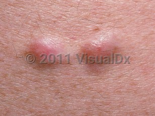
Piloleiomyomas are skin-colored to reddish-brown papules or nodules that are firm to palpation and measure from a few millimeters up to 2 centimeters. They may be solitary or multiple. Solitary lesions tend to be located on the extremities, while multiple agminated or linear lesions favor the torso and may involve more than one site. Up to 90% of affected patients report their lesions to be painful, with most reporting paroxysmal sharp or burning sensations either spontaneously or associated with touch, cold, or emotional stress.
Piloleiomyomas are the subtype most commonly associated with the syndrome of hereditary leiomyomatosis and renal cell cancer (HLRCC, also known as Reed syndrome). These patients have germline heterozygous mutation of the fumarate hydratase gene that is inherited in an autosomal dominant fashion. In this condition, CLs develop by the patients' 20s, uterine leiomyomas develop (in women) by their 30s, and type 2 papillary renal cell cancer develops in the patients' 40s.
Genital CLs tend to be solitary and are more likely nonpainful compared to the other forms of CL. They evolve from dartoic, vulvar, or mammary smooth muscle and may be pedunculated.