Dermatomyositis is a multisystem autoimmune connective tissue disease that is most often characterized by a symmetric proximal extensor inflammatory myopathy, a characteristic violaceous cutaneous eruption, and pathogenic circulating autoantibodies. Dermatomyositis demonstrates a bimodal incidence, with the adult form most commonly seen in individuals aged 45-60 and the juvenile form found in children aged 10-15 years. A 2:1 female-to-male incidence ratio exists in adults.
The distinctive nail findings of dermatomyositis help distinguish it from other connective tissue disorders. Thickened, hyperkeratotic, ragged cuticles and telangiectasias of the proximal nail fold (PNF) are the characteristic nail findings. Some other rare nail findings have been reported, including complete loss of several toenails, red lunulae, and ventral pterygium. The presence of ischemic lesions might be predictive of malignancy in dermatomyositis of adulthood.
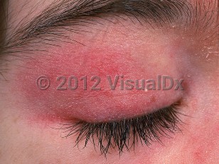
In addition to the nail findings, other cutaneous findings such as periorbital heliotrope rash, atrophic dermal papules of dermatomyositis (ADPDM, formerly called Gottron papules) (slightly atrophic, flat-topped papules over the proximal interphalangeal joints), and poikiloderma should also be present. ADPDM may involve the vicinity of the PNF. Other characteristic features may include flat erythema of the upper back and posterior neck and shoulders (shawl sign) as well as a similarly presenting macular erythema of the anterior neck and upper chest (V sign) that can worsen with ultraviolet exposure. Patients may also have poikiloderma over the lateral hip (holster sign). Muscle involvement affects proximal muscle groups in a symmetric fashion.
Dermatomyositis may be induced by medications, including hydroxyurea, penicillamine, interferon beta, and ipilimumab. Acute onset / flares of dermatomyositis have been reported in association with ingestion of IsaLean, an herbal supplement.
Dermatomyositis - Nail and Distal Digit
See also in: Overview,Cellulitis DDx,External and Internal EyeAlerts and Notices
Important News & Links
Synopsis

Codes
ICD10CM:
M33.10 – Other dermatomyositis, organ involvement unspecified
SNOMEDCT:
396230008 – Dermatomyositis
M33.10 – Other dermatomyositis, organ involvement unspecified
SNOMEDCT:
396230008 – Dermatomyositis
Look For
Subscription Required
Diagnostic Pearls
Subscription Required
Differential Diagnosis & Pitfalls

To perform a comparison, select diagnoses from the classic differential
Subscription Required
Best Tests
Subscription Required
Management Pearls
Subscription Required
Therapy
Subscription Required
Drug Reaction Data
Subscription Required
References
Subscription Required
Last Reviewed:07/25/2019
Last Updated:12/21/2021
Last Updated:12/21/2021
 Patient Information for Dermatomyositis - Nail and Distal Digit
Premium Feature
VisualDx Patient Handouts
Available in the Elite package
- Improve treatment compliance
- Reduce after-hours questions
- Increase patient engagement and satisfaction
- Written in clear, easy-to-understand language. No confusing jargon.
- Available in English and Spanish
- Print out or email directly to your patient
Upgrade Today

Dermatomyositis - Nail and Distal Digit
See also in: Overview,Cellulitis DDx,External and Internal Eye