Epidermoid cyst in Child
See also in: Anogenital,Hair and ScalpAlerts and Notices
Important News & Links
Synopsis

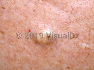
Epidermoid cysts (epidermal cyst, epidermal inclusion cysts, keratin cysts) are frequently incorrectly called sebaceous cysts. One of the most common benign skin tumors in adults, epidermoid cysts are rare in childhood and infancy. These semi-solid cysts are lined by a keratinizing epithelium and filled mostly with macerated keratin, which has a cheese-like consistency and pungent odor. They frequently appear to arise spontaneously. Alternatively, they may result from disruption of follicular structures or by implantation of the epidermis via a penetrating injury. Epidermoid cysts are a feature of several hereditary syndromes, such as Gardner syndrome; cysts are usually multiple and in unusual locations in Gardner syndrome.
Codes
ICD10CM:
L72.0 – Epidermal cyst
SNOMEDCT:
419893006 – Epidermoid cyst
L72.0 – Epidermal cyst
SNOMEDCT:
419893006 – Epidermoid cyst
Look For
Subscription Required
Diagnostic Pearls
Subscription Required
Differential Diagnosis & Pitfalls

To perform a comparison, select diagnoses from the classic differential
Subscription Required
Best Tests
Subscription Required
Management Pearls
Subscription Required
Therapy
Subscription Required
References
Subscription Required
Last Reviewed:05/08/2017
Last Updated:05/08/2017
Last Updated:05/08/2017
 Patient Information for Epidermoid cyst in Child
Premium Feature
VisualDx Patient Handouts
Available in the Elite package
- Improve treatment compliance
- Reduce after-hours questions
- Increase patient engagement and satisfaction
- Written in clear, easy-to-understand language. No confusing jargon.
- Available in English and Spanish
- Print out or email directly to your patient
Upgrade Today

Epidermoid cyst in Child
See also in: Anogenital,Hair and Scalp