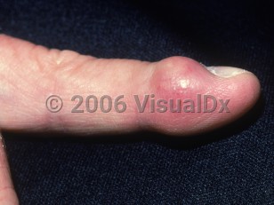
Giant cell tumor of the tendon sheath (GCTTS) is a soft-tissue tumor most commonly found attached to the flexor surfaces of the tendons of the fingers, hands, and wrists. In these locations, the second and third digits are most often involved, particularly near the distal interphalangeal joint. The second most frequent site of occurrence is the ankle-foot region, with toe involvement rare. Individual lesions tend to range from 1-3 cm in diameter and are persistent. GCTTS may occur at any age but is most common in the third to fifth decades. These tumors are typically less frequent in children. The majority of GCTTS present with a history of a gradually enlarging soft-tissue swelling or firm, palpable mass. The tumor may interfere with normal functioning of the joint.
GCTTS has been classified into 2 types. The nodular type is often localized to the hands, has a pseudocapsule, and has a slight predilection for females. The diffuse type is often localized to larger joints and presents with multicentric tumors or satellite lesions without capsules. The diffuse type tends to recur after excision and affects both sexes equally.
GCTTS is thought to result from a reactive inflammatory process. Other hypotheses posit that it arises from the synovial membrane, sesamoid bones, or primitive mesenchymal cells.
Almost all cases are benign, but malignant degeneration has rarely been reported.
Giant cell tumor of tendon sheath
See also in: Nail and Distal DigitAlerts and Notices
Important News & Links
Synopsis

Codes
ICD10CM:
D48.19 – Other specified neoplasm of uncertain behavior of connective and other soft tissue
SNOMEDCT:
95413004 – Nodular tenosynovitis
D48.19 – Other specified neoplasm of uncertain behavior of connective and other soft tissue
SNOMEDCT:
95413004 – Nodular tenosynovitis
Look For
Subscription Required
Diagnostic Pearls
Subscription Required
Differential Diagnosis & Pitfalls

To perform a comparison, select diagnoses from the classic differential
Subscription Required
Best Tests
Subscription Required
Management Pearls
Subscription Required
Therapy
Subscription Required
References
Subscription Required
Last Reviewed:02/26/2019
Last Updated:02/25/2019
Last Updated:02/25/2019
Giant cell tumor of tendon sheath
See also in: Nail and Distal Digit