Epidermal nevi are hamartomas of ectodermal origin. The term encompasses a variety of lesions with various histologic and clinical patterns. Epidermal nevi can be subdivided into keratinocytic and organoid nevi. Keratinocytic epidermal nevus is also known as linear verrucous epidermal nevus.
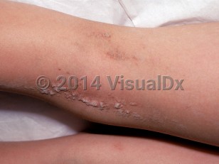
Keratinocytic epidermal nevi are usually recognized at birth but may not become evident until later in childhood, when they present as verrucous skin-colored or hyperpigmented papules coalescing into plaques in a linear or whorled array. The degree of rugosity may vary from one patient to another and increase over time. Mutations in RAS, PIK3CA, and FGFR3 have been reported in these nevi.
Epidermolytic epidermal nevi are a subtype that, if passed on to offspring in germline fashion, yield an infant with bullous congenital ichthyosiform erythroderma eventuating in epidermolytic ichthyosis.
When epidermal nevi are associated with other cutaneous, central nervous system, skeletal, and ocular abnormalities, this is referred to as an epidermal nevus syndrome. Similar mutations are found in the syndromic forms and isolated nevus cases, the more severe presentation perhaps owing to transformation of an earlier progenitor cell yielding change in seemingly unrelated tissues. The genetic loci outlined above are also hot spots for malignancy, and there are an increasing number of reports of internal malignancy associated with extensive epidermal nevi.
Keratinocytic epidermal nevus syndrome, which features epidermal nevi, elevated fibroblast growth factor-23, and hypophosphatemia, has been found to have a genetic mosaicism with the HRAS gene in affected tissues.
Keratinocytic epidermal nevus - Anogenital in
See also in: OverviewAlerts and Notices
Important News & Links
Synopsis

Codes
ICD10CM:
D23.9 – Other benign neoplasm of skin, unspecified
SNOMEDCT:
239107007 – Epidermal nevus
D23.9 – Other benign neoplasm of skin, unspecified
SNOMEDCT:
239107007 – Epidermal nevus
Look For
Subscription Required
Diagnostic Pearls
Subscription Required
Differential Diagnosis & Pitfalls

To perform a comparison, select diagnoses from the classic differential
Subscription Required
Best Tests
Subscription Required
Management Pearls
Subscription Required
Therapy
Subscription Required
References
Subscription Required
Last Reviewed:02/18/2020
Last Updated:03/12/2020
Last Updated:03/12/2020
Keratinocytic epidermal nevus - Anogenital in
See also in: Overview