Keratoacanthomas are most commonly seen in individuals aged 60 years and older with lighter light skin colors and a history of prolonged sun exposure. Men are more commonly affected than women.
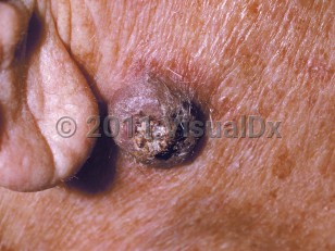
Keratoacanthomas typically present as solitary, crater-shaped nodules measuring a couple centimeters in diameter, often with a central keratin plug on sun-exposed skin.
While solitary keratoacanthomas as described above are the most common presentation, there are variants. The giant type is usually larger and measures between 2-15 cm in diameter, the subungual type occurs underneath the nail, the mucosal type occurs on mucous membranes, and the keratoacanthoma centrifugum marginatum type has a more prominent horizontal growth pattern.
Risk factors for keratoacanthomas include ultraviolet radiation, human papillomavirus infection, immunosuppression, and certain medications. Patients on immunosuppressant medications tend to have more persistent and chronic keratoacanthomas. Patients taking medications such as BRAF inhibitors or hedgehog inhibitors have also been reported to develop keratoacanthomas. In addition, skin injury may be a predisposing factor, and there are reports of keratoacanthomas developing in sites of prior trauma, in surgical scars, after laser resurfacing, and following radiation therapy. In rare cases, keratoacanthomas may develop as part of a syndrome.
- In Ferguson-Smith syndrome, patients develop multiple self-healing keratoacanthomas. It is often seen in younger individuals. It is a genetic condition inherited in an autosomal dominant manner.
- In generalized eruptive keratoacanthomas of Grzybowski, patients develop hundreds to thousands of keratoacanthomas on both the skin and mucous membranes. These keratoacanthomas are frequently chronic and progressive.
- In Muir-Torre syndrome, patients may develop keratoacanthomas along with sebaceous neoplasms and/or gastrointestinal or genitourinary malignancy.
- In familial keratoacanthomas of Witten and Zak, patients develop multiple large and small keratoacanthomas; it is inherited in an autosomal dominant fashion.

 Patient Information for