Langerhans cell histiocytosis (LCH) is a rare (1 in 5 million) clonal histiocytic neoplastic disorder in which immature dendritic histiocytes (CD1a+ / CD207+) accumulate in one or many organs. Activating MAPK pathway mutations, especially in BRAF V600E and MAP2K1, have been shown to be present in an overwhelming majority of patients.
LCH, previously referred to as histiocytosis X, occurs most commonly in White male children aged 1-4 years. Studies in the United States have shown an increased incidence among individuals of Hispanic descent and a lower incidence among Black children. LCH is most often seen in children aged 10 years and younger.
LCH is clinically heterogeneous and classified based on the number of organ systems involved (ie, single-system [SS] or multisystem [MS] LCH). The 4 previously designated clinical variants (Letterer-Siwe disease, Hand-Schüller-Christian syndrome, self-healing reticulohistiocytosis of Hashimoto-Pritzker [congenital self-healing histiocytosis], and eosinophilic granuloma) have largely been abandoned in recent years due to significant overlap between the clinical entities and lack of prognostic relevance.
LCH can affect any organ of the body, including bones, skin, oral mucosa, genital mucosa, nails, bone marrow, lungs, liver, spleen, gastrointestinal tract, lymph nodes, and the central nervous system. The skin is affected in approximately 60% of pediatric LCH cases. The brain, spleen, bone marrow, and liver are considered high-risk organs. Involvement of one or more of these organs is associated with a higher risk of dying from the disease.
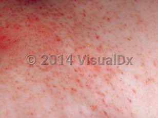
LCH often presents in the first year of life as a rash resembling a seborrheic or eczematous eruption on the scalp, trunk, or diaper area. Ninety percent of patients with skin involvement will also have MS disease. Other findings may include lymphadenopathy, diffuse lung involvement, osteolytic involvement of the mastoid presenting with a picture of otitis media, and gastrointestinal involvement. Failure to thrive may be due to malabsorption, and fevers are often reported. There is wide variation in the clinical spectrum. The congenital form of LCH often involves the skin only and presents with papules, nodules, or ulcers at birth; nodules may be solitary or few.
While lung involvement in the isolated form of the disease is uncommon in children, 25% of patients with MS disease have lung involvement presenting as a nonproductive cough and shortness of breath.
Skull (calvarium) lesions are considered to be a risk factor for central nervous system involvement. These patients may have a higher risk of developing diabetes insipidus and/or neurodegenerative complications. Bone involvement is present in 80% of patients and is often unifocal. While any bone can be affected, the involvement of a bone or bones of the axial skeleton is most common.
Additional disease sequelae can present up to decades after diagnosis, including endocrine (diabetes insipidus, growth delay), skeletal (scoliosis, abnormal dental development, hearing dysfunction), neuropsychological (cerebellar ataxia, learning disabilities), pulmonary, and hepatic involvement (sclerosing cholangitis).
LCH has an unpredictable course and may recur after resolution. LCH has also been associated with the development of leukemias and lymphomas.
Langerhans cell histiocytosis in Infant/Neonate
See also in: AnogenitalAlerts and Notices
Important News & Links
Synopsis

Codes
ICD10CM:
C96.5 – Multifocal and unisystemic Langerhans-cell histiocytosis
C96.6 – Unifocal Langerhans-cell histiocytosis
SNOMEDCT:
65399007 – Langerhans cell histiocytosis
C96.5 – Multifocal and unisystemic Langerhans-cell histiocytosis
C96.6 – Unifocal Langerhans-cell histiocytosis
SNOMEDCT:
65399007 – Langerhans cell histiocytosis
Look For
Subscription Required
Diagnostic Pearls
Subscription Required
Differential Diagnosis & Pitfalls

To perform a comparison, select diagnoses from the classic differential
Subscription Required
Best Tests
Subscription Required
Management Pearls
Subscription Required
Therapy
Subscription Required
References
Subscription Required
Last Reviewed:03/04/2024
Last Updated:03/05/2024
Last Updated:03/05/2024
Langerhans cell histiocytosis in Infant/Neonate
See also in: Anogenital