Contents

SynopsisCodesLook ForDiagnostic PearlsDifferential Diagnosis & PitfallsBest TestsManagement PearlsTherapyDrug Reaction DataReferences

Information for Patients

View all Images (21)

Milia in Child
See also in: External and Internal Eye
Other Resources UpToDate PubMed

Milia in Child

See also in: External and Internal Eye
Contributors: Lowell A. Goldsmith MD, MPH, Craig N. Burkhart MD, Dean Morrell MD
Other Resources UpToDate PubMed

Synopsis

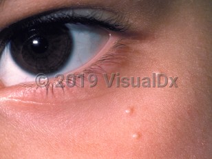
Milia (singular, milium) are minute epidermoid cysts (also known as infundibular cysts) that present as small white or yellow papules, usually on the face of infants and adults although they can also occur in children. They are typically smaller than 3 mm in diameter. Primary milia affect 40%-50% of newborns but may be found in patients of all ages. Secondary milia often occur after injury to the skin, such as from burns or subepidermal blistering disorders (epidermolysis bullosa). Milia have also been known to occur in areas of topical steroid-induced atrophy. Persistent or widespread milia are associated with a number of syndromes. There is no predilection for either sex or for any race or ethnicity.

Codes

ICD10CM:
L72.8 – Other follicular cysts of the skin and subcutaneous tissue

SNOMEDCT:
254679001 – Milia

Look For

Subscription Required

Diagnostic Pearls

Subscription Required

Differential Diagnosis & Pitfalls

Some syndromes associated with milia are as follows:
Blistering disorders that may heal with milia and scarring are as follows:

Best Tests

Subscription Required

Management Pearls

Subscription Required

Therapy

Subscription Required

Drug Reaction Data

Below is a list of drugs with literature evidence indicating an adverse association with this diagnosis. The list is continually updated through ongoing research and new medication approvals. Click on Citations to sort by number of citations or click on Medication to sort the medications alphabetically.

Subscription Required

References

Subscription Required

Last Updated:01/24/2021
Copyright © 2023 VisualDx®. All rights reserved.
Patient Information for Milia in Child
Print E-Mail Images (21)
Contributors: Medical staff writer

Overview

Milia are common non-cancerous (benign) skin findings in people of all ages. Milia formed directly from sloughed-off skin, known as primary milia, are small, fluid-filled cysts usually found on the faces of infants and adults, while lesions formed indirectly, known as secondary milia, are small cysts found within areas of skin affected by another skin condition.

Milia are formed when skin does not slough off normally but instead remains trapped in pockets on the surface of the skin. An individual milium is formed from a hair follicle (pilosebaceous unit) or from a sweat gland (eccrine gland). In primary milia in infants, the oil gland (sebaceous gland) may not be fully developed. Secondary milia often develop after injury or blistering of the skin, which disrupts and clogs the tubes (glandular ducts) leading to the skin surface.

Who’s At Risk

Milia can occur in people of all ages, of any race, and of either sex.

Milia are so common in newborn babies (occurring in up to 50% of them) that they are considered normal.

Secondary milia may appear in affected skin of people with the following:
  • Blistering injury (trauma) to skin, such as poison ivy
  • Burns
  • Blistering skin disorders, such as epidermolysis bullosa or porphyria
  • Following long-term use of topical steroids

Signs & Symptoms

The most common locations for primary milia include:
  • Around the eye (periorbital area) in children and adults
  • Around the nose, especially in infants
The most common locations for secondary milia include:
  • Anywhere on the body, where another skin condition exists
  • On the faces of people who have had a lot of damage from sun exposure
A single lesion (milium) appears as a small (1-2 mm), white-to-yellow, dome-shaped bump on the outer surface of the skin.

Self-Care Guidelines

Although milia are found in the outer (superficial) layers of skin, they are difficult to remove without the proper tools. Do not try to remove them at home as you may leave a scar.

Primary milia found in infants tend to heal on their own within several weeks, though the secondary milia found in older children and adults tend to be long-lasting.

When to Seek Medical Care

See your child's doctor or a dermatologist for evaluation if you notice any new bump on your child's skin.

Treatments

If your doctor diagnoses primary milia in an infant, no treatment is necessary as the condition will go away on its own within a few weeks.

If your child has secondary milia, the doctor will likely treat the other skin condition at that area, if it is still present. Other treatments for milia include:
  • Topical retinoid cream such as tretinoin, tazarotene, or adapalene
  • Removal with a sterile blade (lancet) or scalpel followed by use of a special tool called a comedone extractor
  • A series of fruit acid peels or microdermabrasion procedures at the dermatologist's office

References


Bolognia, Jean L., ed. Dermatology, pp.1722-1723. New York: Mosby, 2003.

Freedberg, Irwin M., ed. Fitzpatrick's Dermatology in General Medicine. 6th ed. pp.601, 604, 780. New York: McGraw-Hill, 2003.
Copyright © 2023 VisualDx®. All rights reserved.
Milia in Child
See also in: External and Internal Eye
A medical illustration showing key findings of Milia (Pediatric) : Cheek, Eyelids, Face, Nose, White color, Tiny papules
Clinical image of Milia - imageId=59031. Click to open in gallery.  caption: 'Few tiny, smooth, white papules near the eye.'
Few tiny, smooth, white papules near the eye.
Copyright © 2023 VisualDx®. All rights reserved.