Morphea is a localized sclerosing condition of the dermis and sometimes the subcutis. It was historically known as localized scleroderma; however, the term morphea is preferred as morphea is distinct from diffuse scleroderma.
The etiology of morphea currently is unknown, although potential exogenous triggers have been suggested, including infection with varicella zoster virus (VZV), Borrelia burgdorferi, or Epstein-Barr virus (EBV); local trauma; sunburn; surgery; and Bacillus Calmette-Guérin (BCG) vaccinations for tuberculosis. The initial event in the pathogenesis is thought to be the induction of vascular damage that leads to activation of the immune system, excessive collagen deposition, and subsequent fibrosis.
In children, morphea is observed in a 2-3:1 female-male ratio, with the mean age of onset ranging from 7-10 years.
Morphea can be divided into the following clinical subtypes: linear, circumscribed / plaque, generalized, pansclerotic, and mixed morphea.
Linear morphea, the most common subtype in childhood, is characterized initially by a linear, indurated plaque. On the limbs, linear morphea is usually unilateral and can lead to limb-length discrepancies and possible limitations in mobility. The "en coup de sabre" variant is named for its location on the paramedian forehead that can cross onto the frontal scalp, with associated alopecia. This subtype can be associated with ocular, neurologic, and odontostomatologic complications. Progressive facial hemiatrophy, also known as Parry-Romberg syndrome, is thought to be a variant of en coup de sabre and involves diffuse unilateral subcutaneous atrophy of the face.
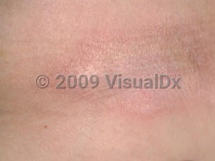
Circumscribed morphea, the second most common subtype in childhood, presents as single or multiple indurated, discrete plaques.
Generalized morphea is characterized by 4 or more plaques larger than 3 cm, involving more than 1 body site.
Pansclerotic morphea is the most severe and rare subtype. It is associated with significant morbidity. Patients present with joint contractures, muscle atrophy, and nonhealing ulcers. There is also an increased risk of squamous cell carcinoma.
Mixed morphea denotes a combination of the features of the aforementioned subtypes.
Extracutaneous manifestations of pediatric morphea are becoming well recognized, as they may affect around one-half to three-quarters of patients. The most commonly affected systems are neurologic, musculoskeletal, ophthalmic, and dental, among others. Contrary to systemic sclerosis, involvement of the renal, cardiac, and pulmonary systems is rare.
Morphea in Child
See also in: External and Internal Eye,Hair and ScalpAlerts and Notices
Important News & Links
Synopsis

Codes
ICD10CM:
L94.0 – Localized scleroderma [morphea]
SNOMEDCT:
201049004 – Morphea
L94.0 – Localized scleroderma [morphea]
SNOMEDCT:
201049004 – Morphea
Look For
Subscription Required
Diagnostic Pearls
Subscription Required
Differential Diagnosis & Pitfalls

To perform a comparison, select diagnoses from the classic differential
Subscription Required
Best Tests
Subscription Required
Management Pearls
Subscription Required
Therapy
Subscription Required
Drug Reaction Data
Subscription Required
References
Subscription Required
Last Reviewed:03/19/2023
Last Updated:04/04/2023
Last Updated:04/04/2023
Morphea in Child
See also in: External and Internal Eye,Hair and Scalp