Oral hairy leukoplakia - Oral Mucosal Lesion
Alerts and Notices
Synopsis

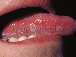
Hairy leukoplakia is a benign mucosal disorder caused by Epstein-Barr virus (EBV) infection and characterized by painless white plaques, typically on the lateral border of the tongue and often bilateral in distribution. The condition is almost always noted in immunocompromised patients.
It was first described in human immunodeficiency virus (HIV)-infected patients but is also seen in patients after organ transplantation. It may be associated with other immunosuppressive states like chemotherapy and hematological malignancies. Very uncommonly, the condition may affect otherwise healthy individuals, and it may be a more transient process in those patients.
The lesions of hairy leukoplakia are asymptomatic and do not scrape off, although they often have a superimposed candidal infection. Lesions develop over weeks to months.
Codes
ICD10CM:
K13.3 – Hairy leukoplakia
SNOMEDCT:
414952002 – Oral hairy leukoplakia
K13.3 – Hairy leukoplakia
SNOMEDCT:
414952002 – Oral hairy leukoplakia
Look For
Subscription Required
Diagnostic Pearls
Subscription Required
Differential Diagnosis & Pitfalls
- Chronic tongue nibbling – This may appear as painless, shaggy white plaques that are bilateral; the buccal mucosa may also be involved. Patients are not usually immunocompromised, although they may coincidentally be so. A biopsy distinguishes between the two, although the patient may be aware of their habit and admit to it.
- Candidiasis – This may appear similar but tends not to be bilaterally symmetrical. The plaques scrape off with some difficulty, leaving a normal or slightly erythematous underlying mucosa.
- Hairy tongue (keratin accumulation on the tips of the filiform papillae) – This generally involves the dorsum of the tongue only, where filiform papillae are located.
- Leukoplakia, especially proliferative verrucous leukoplakia – This may be white and rough-looking but usually does not have a symmetrical distribution. Sharply demarcated margins are characteristic of a preneoplastic process, but biopsy may be the only means to distinguish leukoplakia from hairy leukoplakia.
- Lichen planus – This is often white and bilaterally located in a symmetrical fashion, but it should show interlacing or peripherally radiating white striae in addition to bilateral buccal mucosal involvement.
- Contact stomatitis – These lesions can be caused by a variety of materials, including deteriorating amalgam restorations or artificially flavored cinnamon products. If the lesion contacts a large amalgam (silver) filling when the tongue is at rest, this is a clue. Biopsy may be necessary to confirm the diagnosis. Cinnamon reactions often have an erythematous component, which would be unusual for hairy leukoplakia. A history of use of cinnamon-flavored products is suggestive, and discontinuing the offending agent should result in complete resolution of the lesion within 1 week.
- Uremic stomatitis
Best Tests
Subscription Required
Management Pearls
Subscription Required
Therapy
Subscription Required
References
Subscription Required
Last Reviewed:12/17/2017
Last Updated:07/01/2020