Contents

SynopsisCodesLook ForDiagnostic PearlsDifferential Diagnosis & PitfallsBest TestsManagement PearlsTherapyDrug Reaction DataReferences

Information for Patients

View all Images (27)

Seborrheic dermatitis - Hair and Scalp
See also in: Overview,Anogenital
Other Resources UpToDate PubMed

Seborrheic dermatitis - Hair and Scalp

See also in: Overview,Anogenital
Contributors: Susan Burgin MD, Whitney A. High MD, JD, MEng, Mary Gail Mercurio MD
Other Resources UpToDate PubMed

Synopsis

Seborrheic dermatitis is a common inflammatory papulosquamous condition that affects the sebum-rich areas of the body, including the face, scalp, neck, upper chest, and back. The pathogenesis is not known with certainty, but it may be related to an abnormal immune response to Pityrosporum (Malassezia) yeast, a common skin commensal.

Seborrheic dermatitis presents in infants as a self-limited eruption caused by persistent maternal androgens, or in adults, after adrenarche. Up to 5% of adults are affected by seborrheic dermatitis, and the condition is particularly common after the fifth or sixth decades.

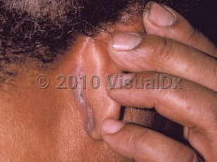
Clinical presentations of seborrheic dermatitis are widely varied, ranging from simple "dandruff" to fulminant rash. There is often dryness, pruritus, erythema, and fine, greasy scaling in characteristic sites, such as the scalp, eyebrows, glabella, nasolabial folds, the beard area, upper chest, external ear canal, posterior ears, eyelid margins (blepharitis), and intertriginous areas. Anogenital involvement has also been reported. One or multiple sites may be involved. In persons with darker skin phototypes, the involved areas may be hypopigmented or hyperpigmented. These pigmentary changes may persist after treatment.

Stress may exacerbate the condition. In immunocompromised persons and those with neurologic conditions, such as Parkinson disease or stroke, seborrheic dermatitis may be severe and recalcitrant.

Since seborrheic dermatitis is such a common disorder, it has been difficult to associate it with specific medications. However, there are some published associations of medications causing, triggering, or exacerbating the condition (see Drug Reaction Data table).

Even with treatment, seborrheic dermatitis tends to be a chronic condition, and remissions and exacerbations are expected. Seborrheic dermatitis is often better in summer months and worse in the winter.

Immunocompromised patient considerations:
  • Seborrheic dermatitis is more common and more severe in persons infected with the human immunodeficiency virus (HIV). It may regress with highly active antiretroviral therapy, but remissions and exacerbations can be expected.
  • Seborrheic dermatitis is also often seen in patients with Parkinson disease. The course is chronic and relapsing and may be difficult to treat.
  • Associated Pityrosporum folliculitis may be seen in immunocompromised patients.

Codes

ICD10CM:
L21.9 – Seborrheic dermatitis, unspecified

SNOMEDCT:
50563003 – Seborrheic dermatitis

Look For

Subscription Required

Diagnostic Pearls

Subscription Required

Differential Diagnosis & Pitfalls

  • Chronic atopic dermatitis patients are often aware of their atopic history, which commonly starts in childhood. More pruritic than psoriasis. The scalp may be involved with pruritic pink plaques with thinner scale, erosions, and excoriations.
  • Psoriasis – Silvery scales sit atop well-demarcated plaques. Seborrheic dermatitis and psoriasis frequently overlap.
  • Tinea capitis – Scale at leading edge of erythema with central clearing. Broken-off hairs or inflammatory boggy plaques (kerion) may be seen. Perform a KOH (potassium hydroxide) preparation of scales and hair.
  • Contact dermatitis – The scalp is initially spared in a contact dermatitis to a contactant that has been applied to the scalp, such as a shampoo. Initially, this will manifest on the skin around the scalp, and only after repeated exposure will the scalp be affected.
  • Dermatomyositis – Scalp involvement manifests with very pruritic and scaly violaceous erythema.
  • Pityriasis amiantacea – Thick adherent scales surround proximal hair shafts. It may be an isolated finding or it may occur in association with inflammatory conditions. Of these, psoriasis and seborrheic dermatitis are most commonly seen.
  • Secondary syphilis – "Moth-eaten" alopecia without scales is seen. Check rapid plasma reagin (RPR) and evaluate for history of primary chancre and systemic symptoms.
  • Crusted scabies – Most often seen in elderly, immunocompromised, or institutionalized patients. Scalp is typically spared; however, there are anecdotal cases of scalp involvement in immunocompromised individuals.

Best Tests

Subscription Required

Management Pearls

Subscription Required

Therapy

Subscription Required

Drug Reaction Data

Below is a list of drugs with literature evidence indicating an adverse association with this diagnosis. The list is continually updated through ongoing research and new medication approvals. Click on Citations to sort by number of citations or click on Medication to sort the medications alphabetically.

Subscription Required

References

Subscription Required

Last Reviewed:02/13/2017
Last Updated:04/05/2021
Copyright © 2023 VisualDx®. All rights reserved.
Patient Information for Seborrheic dermatitis - Hair and Scalp
Print E-Mail Images (27)
Contributors: Medical staff writer
Premium Feature
VisualDx Patient Handouts
Available in the Elite package
  • Improve treatment compliance
  • Reduce after-hours questions
  • Increase patient engagement and satisfaction
  • Written in clear, easy-to-understand language. No confusing jargon.
  • Available in English and Spanish
  • Print out or email directly to your patient
Copyright © 2023 VisualDx®. All rights reserved.
Seborrheic dermatitis - Hair and Scalp
See also in: Overview,Anogenital
A medical illustration showing key findings of Seborrheic dermatitis : Erythema, Fine scaly plaque, Pruritus
Clinical image of Seborrheic dermatitis - imageId=144205. Click to open in gallery.  caption: 'Thin scaly plaque in the post-auricular fold.'
Thin scaly plaque in the post-auricular fold.
Copyright © 2023 VisualDx®. All rights reserved.