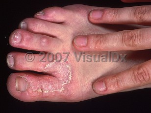
Tinea pedis in Child
See also in: Cellulitis DDxAlerts and Notices
Synopsis

The clinical presentation of tinea pedis may vary. The web spaces and soles are affected most frequently, but the condition may spread to involve the nonplantar surfaces of the foot as well. Interdigital maceration, especially of the lateral toe webs, is commonly seen. Tinea pedis is frequently asymmetric with one foot only being affected or disease being more widespread on one foot than the other. The degree of associated pruritus varies, but most cases are asymptomatic. Trichophyton rubrum may present with a red, scaly, moccasin-like plaque involving the sole. The bullous form of tinea pedis is rare in children and is usually caused by Trichophyton interdigitale (formerly T mentagrophytes var interdigitale). Onychomycosis may be associated.
Interdigital cracking and maceration may act as a portal of entry for pathogens and may predispose to lymphangitis or cellulitis. A dermatophytid reaction (also called an "id reaction") is a hypersensitivity process that can occur secondary to tinea pedis. The condition manifests on the lateral aspects of the fingers and may mimic dyshidrotic dermatitis. This hypersensitivity process will resolve with adequate treatment of the dermatophyte infection.
Immunocompromised patient considerations: In patients with human immunodeficiency virus (HIV) infection and other T-cell disorders, interdigital tinea pedis has been noted to spread to involve the dorsal foot in an extensive manner.
Codes
ICD10CM:B35.3 – Tinea pedis
SNOMEDCT:
6020002 – Tinea pedis
Look For
Subscription Required
Diagnostic Pearls
Subscription Required
Differential Diagnosis & Pitfalls
- Web space erythrasma is typically hyperkeratotic but can be erosive.
- Bullous tinea pedis may be confused with friction blisters or autoimmune blistering disorders.
- Maceration with mixed bacteria (mixed toe web infection)
- Dyshidrotic dermatitis (also called dyshidrotic eczema or pompholyx)
- Contact dermatitis (irritant, allergic)
- Psoriasis
- Juvenile plantar dermatosis
- Palmoplantar keratoderma
- Pitted keratolysis – A frequently overlooked condition that causes plantar pits and malodorous feet. Its cause is bacterial, and it responds to topical antibacterials such as clindamycin solution and aluminum chloride 20% solution for drying.
Best Tests
Subscription Required
Management Pearls
Subscription Required
Therapy
Subscription Required
References
Subscription Required
Last Reviewed:08/15/2019
Last Updated:12/09/2020


Overview
Athlete's foot (tinea pedis), also known as ringworm of the foot, is a surface (superficial) fungal infection of the skin of the foot. Though it is not commonly found in children, athlete's foot is the most common fungal disease in humans.Athlete's foot may be passed to humans by direct contact with infected people, infected animals, contaminated objects (such as towels or locker room floors), or the soil.
Who’s At Risk
Athlete's foot may occur in people of all ages, of all races, and of both sexes. Young children rarely develop athlete's foot, though it is frequently seen in teens (adolescents) and adults. In addition, athlete's foot is more common in males than in females.Some conditions make athlete's foot infections more likely to occur:
- Living in warm, humid climates
- Using public or community pools or showers
- Wearing tight, non-ventilated footwear
- Sweating profusely
- Having diabetes or a weakened immune system
Signs & Symptoms
The most common locations for athlete's foot include:- Spaces (webs) between the toes, especially between the 4th and 5th toes and between the 3rd and 4th toes
- Soles of the feet
- Tops of the feet (very unusual in children)
- Between the toes (the interdigital spaces), athlete's foot may appear as inflamed, scaly, and soggy tissue. Splitting of the skin, called fissures, may be present between or under the toes. This form of athlete's foot tends to be quite itchy.
- On the sole of the foot (the plantar surface), athlete's foot may appear as pink-to-red skin with scales ranging from mild to widespread (diffuse).
- On the top of the foot, athlete's foot appears as one or more red, scaly patches ranging in size from 1-5 cm. The border of the affected skin may be raised and may contain bumps, blisters, or scabs. Often, the central portion of the lesion is clear, leading to a ring-like shape and the descriptive (but inaccurate) name "ringworm."
- Another type of tinea infection, called bullous tinea pedis, appears as painful and itchy blisters on the arch (instep) and/or the ball of the foot.
- The most severe form of the infection, called ulcerative tinea pedis, appears as painful blisters, pus-filled bumps (pustules), and shallow ulcers. These lesions are especially common between the toes but may involve the entire sole. Because of the numerous breaks in the skin, lesions commonly become infected with bacteria. Ulcerative tinea pedis occurs most frequently in people with diabetes and others with weakened immune systems.
Self-Care Guidelines
If you suspect that your child has athlete's foot, you might try one of the following over-the-counter antifungal creams or lotions:- Terbinafine
- Clotrimazole
- Miconazole
In addition, try to keep your child's feet dry, creating conditions where the dermatophyte cannot live and grow. Have your child try the following:
- Wash his or her feet daily and dry them carefully, even using a hair dryer (on low setting) if necessary.
- Use a separate towel for the feet, and do not share this towel with anyone else.
- Wear socks made of cotton or wool, and change them once or twice a day, or even more often if they become damp.
- Avoid wearing shoes made of synthetic materials such as rubber or vinyl.
- Wear sandals as often as possible.
- Apply antifungal powder to the feet and inside the shoes every day.
- Wear protective footwear in locker rooms and public or community pools and showers.
When to Seek Medical Care
If the lesions do not improve after 2 weeks of applying over-the-counter antifungal creams, or if they are exceptionally itchy or painful, see your child's doctor for an evaluation. If your child has blisters, pustules, and/or ulcers on the feet, see a doctor as soon as possible.Treatments
To confirm the diagnosis of athlete's foot, your child's doctor might scrape some surface skin material (scales) onto a glass slide and examine them under a microscope. This procedure, called a KOH (potassium hydroxide) preparation, allows the doctor to look for tell-tale signs of fungal infection.Once the diagnosis of athlete's foot has been confirmed, the physician will probably start treatment with an antifungal medication. Most infections can be treated with topical creams and lotions, including:
- Over-the-counter preparations such as terbinafine, clotrimazole, or miconazole
- Prescription-strength creams such as econazole, oxiconazole, ciclopirox, ketoconazole, sulconazole, naftifine, or butenafine
- Compounds containing urea, lactic acid, or salicylic acid to help dissolve the scale and allow the antifungal cream to penetrate better into the skin
- Solutions containing aluminum chloride, which reduce sweating of the foot
- Antibiotic creams to prevent or treat bacterial infections, if present
- Griseofulvin
- Terbinafine
References
Bolognia, Jean L., ed. Dermatology, pp.1174-1185. New York: Mosby, 2003.
Freedberg, Irwin M., ed. Fitzpatrick's Dermatology in General Medicine. 6th ed. pp.1251, 2000-2001, 2337, 2340-2041, 2446-2447. New York: McGraw-Hill, 2003.
Tinea pedis in Child
See also in: Cellulitis DDx