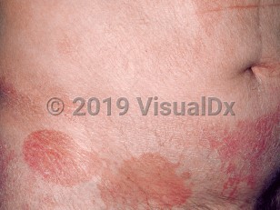
Cutaneous T-cell lymphomas (CTCLs) are a heterogeneous group of neoplasms that vary considerably in their clinical presentation, histology, immunophenotype, genetics, and prognosis. Mycosis fungoides (MF) and its variants (folliculotropic mycosis fungoides, pagetoid reticulosis, and granulomatous slack skin), Sézary syndrome, lymphomatoid papulosis, and cutaneous anaplastic large cell lymphoma make up 90% of all CTCL cases. CTCLs are seen most often in the elderly, but they can occur in patients of all ages. The definitive diagnosis of CTCL may require large or multiple biopsies as well as specialized testing of the biopsy specimens. Typing of the CTCL and staging are important to determine the extent of disease and treatment strategy.
Nail changes are common in patients with CTCL, especially those with Sézary syndrome, and typically affect more than one nail. In many cases, changes affect all 20 nails. At times, the nail changes are due to treatments of CTCL and not to the disease itself. For example, Beau lines have been observed with chemotherapy, and melanonychia was seen with psoralen plus ultraviolet A (PUVA) therapy.
In a retrospective study of 83 patients with Sézary syndrome, 36 patients (43.4%) were found to have nail changes. Nail thickening, nonspecific nail dystrophy, nail plate yellowing, and subungual hyperkeratosis were among the most common nail findings (42%-58%). Other nail changes were onycholysis, nail plate ridging, and splinter hemorrhages, seen in 17%-19% of patients. Beau lines and onychomadesis were seen in 8% of patients. In another study, 52.9% (45 of 85) of patients with Sézary syndrome who had keratoderma had positive potassium hydroxide (KOH) tests under microscopy on skin scrapings. Therefore, some of these nail changes may be attributable to onychomycosis. Paronychia was frequently reported in another study of 19 patients with Sézary syndrome (63.2%).
Small case series and case reports have described nail discoloration and thickening, onycholysis, crumbling, subungual hyperkeratosis, anonychia, onychomadesis, and splinter hemorrhages in patients with MF. In a retrospective study of 60 patients with biopsy-proven MF, 18 (30%) had nail changes. The most common nail findings were longitudinal ridging, nail plate thickening, nail fragility, and leukonychia.
Cutaneous T-cell lymphoma - Nail and Distal Digit
See also in: OverviewAlerts and Notices
Important News & Links
Synopsis

Codes
ICD10CM:
C84.A0 – Cutaneous T-cell lymphoma, unspecified, unspecified site
SNOMEDCT:
400122007 – Primary cutaneous T-cell lymphoma
C84.A0 – Cutaneous T-cell lymphoma, unspecified, unspecified site
SNOMEDCT:
400122007 – Primary cutaneous T-cell lymphoma
Look For
Subscription Required
Diagnostic Pearls
Subscription Required
Differential Diagnosis & Pitfalls

To perform a comparison, select diagnoses from the classic differential
Subscription Required
Best Tests
Subscription Required
Management Pearls
Subscription Required
Therapy
Subscription Required
Drug Reaction Data
Subscription Required
References
Subscription Required
Last Reviewed:07/15/2020
Last Updated:02/14/2022
Last Updated:02/14/2022
 Patient Information for Cutaneous T-cell lymphoma - Nail and Distal Digit
Premium Feature
VisualDx Patient Handouts
Available in the Elite package
- Improve treatment compliance
- Reduce after-hours questions
- Increase patient engagement and satisfaction
- Written in clear, easy-to-understand language. No confusing jargon.
- Available in English and Spanish
- Print out or email directly to your patient
Upgrade Today

Cutaneous T-cell lymphoma - Nail and Distal Digit
See also in: Overview