Acroosteolysis describes the occurrence of destructive changes of the distal phalanges. Progressive destruction of the bone produces peg-shaped phalanges, and fingertips may appear bulbous. Acroosteolysis is frequently in the presence of distal digital ischemia, digital calcinosis, and sensory neuropathy.
Radiologic categorization of acroosteolysis is based on the resorption pattern seen in the distal phalanx. The transverse type is associated with perpendicular lines of resorption on the distal phalangeal shafts, and the longitudinal type, which is more common, is associated with resorption of the distal phalangeal terminal tufts.
There are multiple occupational, autoimmune, infectious, hormonal / metabolic, and genetic causes of acroosteolysis.
Occupational / behavioral causes include polyvinyl chloride (PVC) exposure, phenytoin exposure in utero, repeated chronic trauma to the phalanges, carpal tunnel syndrome, and thermal injury (including burns and frostbite).
Individuals cleaning or inspecting autoclaves for polymerization of vinyl chloride to PVC have a high incidence of the development of acroosteolysis. The disease is also characterized by Raynaud phenomenon, sclerodactyly, and papular fibrotic lesions on the wrists and dorsa of the hands. Inhalation of the vinyl chloride monomer is the method of exposure.
Autoimmune causes include systemic sclerosis, psoriatic arthritis, systemic lupus erythematosus, granulomatosis with polyangiitis, mixed connective tissue disease, juvenile chronic arthritis, and Raynaud phenomenon. Acroosteolysis can also be seen in sarcoidosis. Acroosteolysis has been associated with digital ischemia and digital ulcers in patients with systemic sclerosis, leading to the theory that acroosteolysis arises due to vascular insufficiency. Additionally, it is associated with calcinosis as part of CREST syndrome. Of those individuals with systemic sclerosis, 20%-25% are estimated to have acroosteolysis.
Leprosy may cause acroosteolysis. This is usually seen as a result of peripheral alteration in sensation secondary to peripheral neuropathy or nerve trunk infiltration.
Acroosteolysis can also be seen in hyperparathyroidism and diabetes mellitus type 1. Genetic causes of acroosteolysis include NOG mutations that encode noggin (a polypeptide that facilitates connective tissue development), Penttinen syndrome, laminopathies, pachydermoperiostosis, hereditary sensory neuropathies, Hajdu-Cheney syndrome, Gaucher disease, intestinal lymphangiectasia, and pyknodysostosis.
Pediatric Considerations: Of note, psoriatic arthritis and systemic sclerosis are the most common rheumatologic causes of acroosteolysis in pediatric patients. Sympathetic denervation due to spinal dysraphism can also lead to lower extremity acroosteolysis in children.
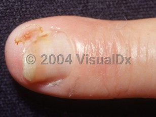
Acroosteolysis - Nail and Distal Digit
Alerts and Notices
Important News & Links
Synopsis

Codes
ICD10CM:
M89.549 – Osteolysis, unspecified hand
SNOMEDCT:
27201004 – Acroosteolysis
M89.549 – Osteolysis, unspecified hand
SNOMEDCT:
27201004 – Acroosteolysis
Look For
Subscription Required
Diagnostic Pearls
Subscription Required
Differential Diagnosis & Pitfalls

To perform a comparison, select diagnoses from the classic differential
Subscription Required
Best Tests
Subscription Required
Management Pearls
Subscription Required
Therapy
Subscription Required
References
Subscription Required
Last Reviewed:01/11/2023
Last Updated:02/02/2023
Last Updated:02/02/2023