Contents

SynopsisCodesLook ForDiagnostic PearlsDifferential Diagnosis & PitfallsBest TestsManagement PearlsTherapyReferences

View all Images (20)

Birt-Hogg-Dubé syndrome
Other Resources UpToDate PubMed

Birt-Hogg-Dubé syndrome

Contributors: Nicole S. Gunasekera BA, William M. Lin MD, Susan Burgin MD, Lowell A. Goldsmith MD, MPH, Ryan R. Walsh MD, Joshua J. Jarvis MD, Alastair Moore MD
Other Resources UpToDate PubMed

Synopsis

Birt-Hogg-Dubé (BHD) syndrome is a rare autosomal dominant genodermatosis known to affect approximately 200 families worldwide. It is caused by a germline mutation in the FLCN gene on chromosome 17p11.2, which encodes the protein folliculin. Disease manifestation varies among those affected but can involve the skin, lungs, and kidneys.

The European Birt-Hogg-Dubé consortium proposed guidelines for the diagnosis as fulfilling either one major or two minor criteria.

Major criteria:     
  1. At least five fibrofolliculomas or trichodiscomas, at least one histologically confirmed, of adult onset.
  2. Pathogenic FLCN germline mutation.  
Minor criteria:
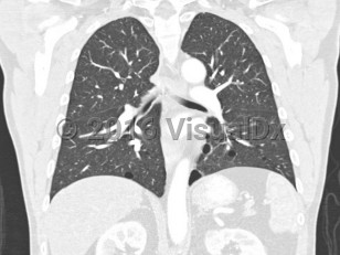
  1. Multiple lung cysts: bilateral basally located lung cysts with no other apparent cause, with or without spontaneous primary pneumothorax.
  2. Renal cancer: early onset (age younger than 50 years) or multifocal or bilateral renal cancer or renal cancer of mixed chromophobe and oncocytic histology.
  3. A first degree relative with BHD.  
Fibrofolliculomas are benign hamartomas of the hair follicle and are the most common manifestation of BHD. Some believe that the fibrofolliculomas and trichodiscomas listed in the major criteria are the same entity on a histologic spectrum. They generally appear in the second or third decade of life and most commonly affect the face and upper torso. In many patients, new fibrofolliculomas develop over time, and existing lesions may grow in size. Acrochordons may be skin-colored or whitish. When acrochordons are biopsied, histopathologic findings of fibrofolliculoma may be seen.

Approximately 80% of patients develop pulmonary cysts, which can be numerous and bilateral. They commonly affect the base of the lungs. They typically develop between the ages of 30 and 40 but have been described from ages 20-85. BHD patients are at 50-fold increased risk of spontaneous pneumothorax, with approximately 24% of those with pulmonary cysts developing spontaneous pneumothoraces at an average age of 38 years (range: 22-71 years). FLCN-mutation carriers have developed pneumothoraces as early as age 7.

Renal tumors occur in approximately 12%-34% of BHD patients at a mean age of 50.4 years (range: 30-70 years). Patients have a 7-fold increased risk of renal tumors compared to the general population. Renal tumors can be bilateral, unilateral, or multifocal. The most common types of renal tumors in this patient population are chromophobe tumors and hybrid chromophobe / oncocytic tumors; however, clear cell carcinoma, papillary carcinoma, and mixed-type carcinoma may also occur.

Codes

ICD10CM:
Q87.89 – Other specified congenital malformation syndromes, not elsewhere classified

SNOMEDCT:
110985001 – Multiple fibrofolliculomas

Look For

Subscription Required

Diagnostic Pearls

Subscription Required

Differential Diagnosis & Pitfalls

To perform a comparison, select diagnoses from the classic differential

Subscription Required

Best Tests

Subscription Required

Management Pearls

Subscription Required

Therapy

Subscription Required

References

Subscription Required

Last Updated:11/29/2023
Copyright © 2024 VisualDx®. All rights reserved.
Birt-Hogg-Dubé syndrome
A medical illustration showing key findings of Birt-Hogg-Dubé syndrome : Smooth papule
Imaging Studies image of Birt-Hogg-Dubé syndrome - imageId=8346944. Click to open in gallery.  caption: '<span>Coronal 2 mm slice thickness  CT image viewed in lung windows. Multiple lung cysts with a basilar  predominance, with the largest cyst located 	in the lower lobes. Many of the  cysts are in a subpleural position. Note thin cyst walls (<1), and lack  of internal structure within the cysts.</span>'
Coronal 2 mm slice thickness CT image viewed in lung windows. Multiple lung cysts with a basilar predominance, with the largest cyst located in the lower lobes. Many of the cysts are in a subpleural position. Note thin cyst walls (<1), and lack of internal structure within the cysts.
Copyright © 2024 VisualDx®. All rights reserved.