Bullous impetigo (pediatric) - Anogenital in
See also in: OverviewAlerts and Notices
Synopsis

Bullous impetigo is a localized form of staphylococcal scalded skin syndrome caused by exfoliative toxins (A and B) released by (phage group II) Staphylococcus aureus. These toxins cleave desmoglein 1, resulting in superficial blisters locally at the site of infection. It is primarily seen in children, especially infants, who lack antibodies against exfoliative toxins, and only rarely occurs in teenagers or young adults. Infection is spread by direct contact with colonized or infected individuals. Staphylococcus aureus often colonizes the nares, umbilicus, nails, and eyes; approximately 5% of S aureus has exfoliative toxin.
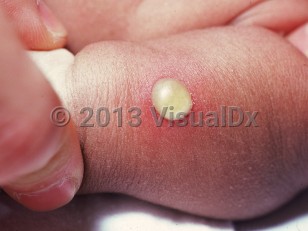
Bullous impetigo initially presents as flaccid bullae, which then rupture, leaving round erosions that become crusted. Constitutional symptoms and fever are rare and mild, if they occur. The disease commonly affects the intertriginous areas, face, and extremities.
Outbreaks tend to occur during the summer months and in humid climates. Full resolution typically occurs within 2-6 weeks. Rare progression to staphylococcal scalded skin syndrome can occur.
In neonates, the infection often presents in the first 2 weeks of life. Sometimes, bullous impetigo may result in serious infections like osteomyelitis, septic arthritis, pneumonia, and septicemia.
Codes
ICD10CM:L01.03 – Bullous impetigo
SNOMEDCT:
399183005 – Impetigo bullosa
Look For
Subscription Required
Diagnostic Pearls
Subscription Required
Differential Diagnosis & Pitfalls
- Varicella – Typically features polymorphic lesions in various stages: macules, vesicles, pustules, and crusting. Face, trunk, and proximal extremities are involved.
- Tinea cruris or tinea corporis (bullous)
- Stevens-Johnson syndrome – Characteristic target lesions (necrotic center surrounded by erythema and edema [pallor]) along with hemorrhagic crusting of the lips and conjunctiva. There may be associated systemic symptoms.
- Erythema toxicum neonatorum – Usually noticed in the first few days of life. The lesions are erythematous macules or urticarial plaques topped with a 1-2 mm papule or pustule that spontaneously resolves within 1-2 days.
- Transient neonatal pustular melanosis – Presents in the immediate postnatal period and is characterized by vesiculopustules without associated erythema. The pustules rupture easily, leaving behind hyperpigmented macules that may be surrounded by a characteristic collarette of scale.
- Staphylococcal scalded skin syndrome
- Bullous fixed drug eruption – Well-demarcated, circular or oval, erythematous patches that recur in the same site (usually lips and trunk) each time the offending drug is administered. Lesions characteristically heal with hyperpigmentation.
- Herpes simplex virus (HSV) infection – Tiny grouped vesicles on an erythematous base that rupture to form polycyclic erosions. Prodromal symptoms are usually present. The skin of the face and hands is commonly affected.
- Neonatal herpes simplex virus infection
- Bullous insect bite reactions – Linear, irregular streaks of dermatitis with vesiculation at the site of bite, often with a "kissing pattern."
- Scabies – Pruritic, erythematous papules and vesiculopustules on the intertriginous areas, face, genitalia, and palms and soles. Burrows may be present in the finger web spaces, flexor aspects of the wrists, axillae, umbilicus, nipples, buttocks, and penis.
- Cutaneous candidiasis affects the intertriginous areas, especially the groin and neck, in the form of confluent, erythematous patches with multiple small satellite pustules. Potassium hydroxide (KOH) test from a pustular lesion reveals budding spores and pseudohyphae that confirm Candida infection.
- Chronic bullous dermatosis of childhood – Tense (subepidermal) blisters in the groin, lower abdomen, back, and perioral region. The characteristic rosette-like vesicles resemble a cluster of pearls surrounding a central healing bulla.
- Contact dermatitis
- Poison ivy or oak dermatitis
- Burns (see thermal or electrical burn, burn marks of child abuse)
- Bullous pemphigoid
- Epidermolysis bullosa simplex
- Sexual abuse – Cases have been reported of bullous impetigo affecting the vulvar region, leading to confusion with possible sexual abuse.
Best Tests
Subscription Required
Management Pearls
Subscription Required
Therapy
Subscription Required
References
Subscription Required
Last Reviewed:03/22/2022
Last Updated:05/05/2022


Overview
Impetigo is a common and contagious bacterial skin infection that is usually a minor problem, but sometimes complications may occur that require treatment. Complications related to impetigo can include deeper skin infection (cellulitis), infections of the brain, and kidney inflammation. Impetigo often starts with a cut or break in the skin that allows bacteria to enter. Impetigo is usually caused by "staph" (Staphylococcus) or "strep" (Streptococcus) bacteria. Impetigo can be further classified into 2 types: bullous and nonbullous.- Nonbullous impetigo accounts for 70% of all cases and appears as tiny fluid-filled blisters that develop into honey-colored, crusty lesions. Generally they do not cause any pain or redness to the surrounding skin.
- Bullous impetigo appears as larger clear blisters filled with fluid. When these blisters break, they may leave a scale behind. Bullous impetigo is primarily seen in infants and children. It is less common in teenagers and young adults.
Who’s At Risk
Bullous impetigo is more commonly seen in infants and usually develops on the face, buttocks, and diaper area. Infants are at a greater risk for these infections because their immune systems are not fully developed.Impetigo is very common in children, affecting up to 10% of children who come to a pediatric clinic. Children up to 6 years old are most likely to be infected.
Those who live in a warm, humid climate are more often affected. Insect bites, crowded living conditions, and poor skin cleansing increase the risk of infection. Impetigo may spread easily through schools, daycare centers, and nurseries. Participating in sports requiring skin-to-skin contact, having a weak immune system, or having a chronic skin problem such as eczema can also increase your child's risk of getting impetigo. Lesions on the neck and scalp may occur with head lice (pediculosis capitis).
Signs & Symptoms
- Painless blisters (about an inch or less) occur that may break easily.
- These often spread to the face, trunk, arms, or legs.
- The child feels generally well (unless it is severe).
Moderate – There are over 10 spots, and several small skin areas are affected.
Severe – There are many lesions, large areas of skin are affected, and/or the child feels ill with fever, diarrhea, or weakness.
Self-Care Guidelines
Prevention:- Keep the skin clean with soap and water.
- Treat cuts, scrapes, and insect bites by cleaning with soap and water and covering the area if possible.
- Gently wash the area with a mild soap and water twice or more daily and cover with gauze or a non-stick dressing if possible.
- Apply an over-the-counter antibiotic ointment after washing the skin 3-4 times daily. Wash hands after application, or wear gloves to apply.
- To remove crusts, soak with a vinegar solution (1 tablespoon of white vinegar to a pint of water) for 15-20 minutes.
- Wash clothing, towels, and bedding daily, and do not share these with others.
- Wash hands frequently, patient should try not to touch the affected areas, and keep fingernails trimmed.
- Keep your child home until scabs or open areas have healed.
When to Seek Medical Care
See your child's doctor for any infection that does not improve. See the doctor immediately for moderate to severe infection or if your child has a fever or severe pain.If your child is currently being treated for a skin infection that has not improved after 2-3 days of antibiotics, return to the child's doctor.
Community-associated methicillin-resistant Staphylococcus aureus (CA-MRSA) is a strain of "staph" bacteria resistant to antibiotics in the penicillin family. CA-MRSA previously infected only a few people, such as health care workers and IV drug users. However, CA-MRSA is now a common cause of skin infections in the general population. While CA-MRSA bacteria are resistant to penicillin and penicillin-related antibiotics, most staph infections with CA-MRSA can be easily treated by health care practitioners using local skin care and commonly available non-penicillin-family antibiotics. Rarely, CA-MRSA can cause serious or deeper skin infections. Staph infections typically start as small red bumps or pus-filled bumps, which can rapidly turn into deep, painful sores. If you see a red bump or pus-filled bump on your child's skin that is worsening or showing any signs of infection (ie, the area becomes increasingly painful, red, or swollen), see the child's doctor right away. Many people believe incorrectly that these bumps are the result of a spider bite when they arrive at the doctor's office. Your doctor may need to test (culture) infected skin for MRSA before starting antibiotics. If your child has a skin problem that resembles a CA-MRSA infection or a culture that is positive for MRSA, the doctor may need to provide local skin care and prescribe oral antibiotics. To prevent spread of infection to others, infected wounds, hands, and other exposed body areas should be kept clean and wounds should be covered during therapy.
Treatments
In addition to the treatments for mild impetigo already mentioned, the doctor may prescribe:- Topical antibiotics (eg, mupirocin or retapamulin), or
- Oral antibiotics (eg, a cephalosporin, clindamycin, or erythromycin).
Bullous impetigo (pediatric) - Anogenital in
See also in: Overview