Linear IgA bullous disease (LABD) of childhood, also known as chronic bullous dermatosis of childhood (CBDC) and chronic bullous disease, is recognized as the childhood counterpart to linear IgA bullous dermatosis. LABD is an immunobullous dermatosis due to immunoglobulin A (IgA) autoantibodies against antigens in the basement membrane that cause subepidermal blister formation. It has clinical similarities to childhood bullous pemphigoid, and some believe them to be on a clinical spectrum of disease.
Childhood LABD usually presents around age 5 years, but it has been seen in neonates up to children aged 10 years. There is a 3:2 female-to-male predominance. This condition is usually self-limited over several months, up to 2-4 years, following a less chronic course than childhood pemphigoid, pemphigus, or dermatitis herpetiformis. While the majority of cases resolve before puberty, a small proportion of patients will have recurrences into adulthood.
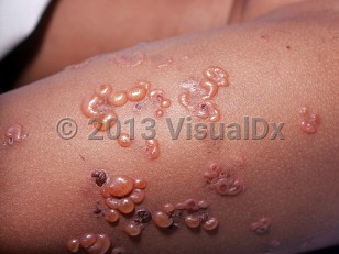
Classically, the lesions appear as clusters or rings of tense bullae in the perioral and perineal regions. Lesions tend to burn or itch, with the blistering becoming less severe with time. The bullae heal with pigmentary changes and infrequently with scarring. Mucosal involvement is rarer in children than in adults.
Drug-induced LABD may be seen in childhood. Clinical presentation is typically indistinguishable from the classic idiopathic form with the exception that large erosions may be more commonly seen in the drug-induced compared with the idiopathic form. Localized or widespread macular erythema may be seen in association. A number of drugs have been associated with LABD, most commonly a variety of antibiotics (eg, trimethoprim-sulfamethoxazole, amoxicillin-clavulanate, and amoxicillin alone), NSAIDs, and cephalosporins.
Most patients with childhood LABD have IgA1 antibodies to a 97-kDa and a 120-kDa fragment of the extracellular portion of bullous pemphigoid antigen 2 (BP180 / type XVII collagen), typically the 15 collagenous domain and less frequently the NC16A epitope. Reactivity to collagen VII, laminin-332, and laminin gamma-1 have also been described.
Linear IgA bullous dermatosis of childhood in Infant/Neonate
Alerts and Notices
Important News & Links
Synopsis

Codes
ICD10CM:
L13.8 – Other specified bullous disorders
SNOMEDCT:
109250009 – Chronic bullous dermatosis of childhood
L13.8 – Other specified bullous disorders
SNOMEDCT:
109250009 – Chronic bullous dermatosis of childhood
Look For
Subscription Required
Diagnostic Pearls
Subscription Required
Differential Diagnosis & Pitfalls

To perform a comparison, select diagnoses from the classic differential
Subscription Required
Best Tests
Subscription Required
Management Pearls
Subscription Required
Therapy
Subscription Required
Drug Reaction Data
Subscription Required
References
Subscription Required
Last Reviewed:02/12/2020
Last Updated:03/31/2024
Last Updated:03/31/2024
Linear IgA bullous dermatosis of childhood in Infant/Neonate