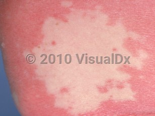
Pityriasis rubra pilaris (PRP) is characterized by an acute cutaneous eruption that is usually accompanied by pruritus and pain. There are both acquired and heritable forms. Classic cutaneous lesions include follicular keratotic papules on an erythematous base coalescing to form large, orange-red plaques with characteristic islands of sparing. PRP commonly begins on the scalp and rapidly spreads in a craniocaudal direction, and it has the potential to quickly progress to erythroderma over several weeks' time. Additional features include an orange-red or bright yellow palmoplantar keratoderma. PRP can be classified into 6 clinical types. The clinical types have been suggested to predict response to therapy and disease course, but this has not been validated.
- Type I (classic adult type, 55% of cases) – Occurs in adults. It is the most common subtype and may clear without therapy within 3-5 years. Relapses are uncommon.
- Type II (atypical adult type, 5% of cases) – A rare subtype seen in adults. It is characterized by more ichthyosiform (plate-like) scale, increased hair loss, and a chronic and severe course.
- Type III (classic juvenile type, 10% of cases) – Occurs in children and adolescents. It resembles type I PRP clinically, although erythroderma is less common. Many cases resolve in less than 3 years, but some cases persist longer.
- Type IV (circumscribed juvenile type, 25% of cases) – Most common subtype in children and adolescents. It is characterized by focal lesions on the extensor surfaces of extremities, over the Achilles, and palmoplantar keratoderma. The clinical course is variable.
- Type V (atypical juvenile type, 5% of cases) – Typically occurs in the first 2 years of life. It is the most common familial form of PRP and may be associated with CARD14 gene variations, although of note, individuals with CARD14 gene variations (also termed CARD14-associated papulosquamous eruption), may present with features of atypical PRP, classical PRP, or psoriasis.
- Type VI (HIV-associated type, < 1% of cases) – Occurs in HIV-infected individuals. It may resemble type I PRP or may be part of a follicular occlusion tetrad that includes acne conglobata, hidradenitis suppurativa, and lichen spinulosus.
The etiology of PRP has not been clearly defined. The onset of disease has been associated with vaccines, infection, malignancy, ultraviolet (UV) exposure, and minor skin trauma. Medications have been implicated as causative in a small number of patients, but more studies are needed. Potential culprits include tyrosine kinase inhibitors (ponatinib, imatinib, sorafenib), topical Toll-like receptor 7 (TLR7) agonists (imiquimod), phosphoinositide 3-kinase inhibitors (ofatumumab and idelalisib), antiviral medications (telaprevir, sofosbuvir), and less commonly, programmed cell death protein 1 (PD-1) inhibitors (pembrolizumab), vascular endothelial growth factor (VEGF) inhibitors (bevacizumab), statins (simvastatin), insulin, and angiotensin-converting enzyme (ACE) inhibitors (ramipril). While infliximab can be an effective treatment for PRP, there is one case report of infliximab-induced PRP.
While there are reports of heritable forms, the large majority of PRP cases are acquired and without sex predilection. The incidence of the acquired form occurs in 2 slight peaks: during the first and second decades and during the sixth decade, although it may present at any age.