Pyoderma gangrenosum in Adult
See also in: Cellulitis DDx,AnogenitalAlerts and Notices
Important News & Links
Synopsis

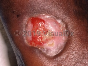
Pyoderma gangrenosum (PG) is an inflammatory, noninfectious, ulcerative neutrophilic skin disease of uncertain etiology commonly misdiagnosed as an aggressive skin infection. Pustules form and give way to ulcers with a necrotic, undermined margin. PG can affect any age and take on a number of differing clinical presentations. PG can have either an acute or chronic course and result in extensive scarring (which can be keloidal or have dyspigmentation, especially in patients with darker skin types). There is no predilection for sex or any population. The disease occurs most often in middle-aged adults.
The 2 primary variants are a classic ulcerative form, which often involves the lower extremities, and a vesicobullous form, which is more superficial and tends to occur on the upper extremities, including hands. Fever, toxicity, and pain can be associated with the onset of PG. Rarely, PG can involve the eyes as well. Extracutaneous manifestations may take the form of sterile neutrophilic abscesses, such as in the lungs, heart, gastrointestinal tract, liver, eyes, central nervous system, and lymphatic tissue.
Necrotizing neutrophilic dermatosis describes a subset of patients with severe acute febrile neutrophilic dermatosis (Sweet syndrome) or PG who develop, in addition to their cutaneous disease, fever, leukocytosis (or a leukemoid reaction), and features of shock. Skin pain is a prominent symptom. Additionally, soft tissues underlying areas of skin involvement may be affected with neutrophilic infiltrates and necrosis.
Though the exact cause is unknown, neutrophil dysfunction, inflammation, and genetics are all thought to play a role. Additionally, PG has associations with a number of systemic illnesses. In about 50% of cases, there is an association between PG and systemic diseases such as ulcerative colitis, Crohn disease, arthritis, myeloma, leukemia, monoclonal gammopathy, granulomatosis with polyangiitis (formerly known as Wegener granulomatosis), collagen vascular disease, metabolic syndrome, and Behçet disease, among other disorders, including genetic conditions (see Look For). Surgery by itself can be a precipitating cause. Levamisole-contaminated cocaine has been associated with PG lesions ranging from vesicopustules to bullae to larger ulcers; most patients demonstrated positivity for antiphospholipid or anticardiolipin antibodies.
PG tends to be self-limited. First-line therapies are widely accepted, while alternative therapeutic recommendations are largely based on anecdotal evidence. Surgical intervention is a common exacerbating factor because PG demonstrates pathergy, a phenomenon by which skin trauma can lead to worsening disease.
Codes
ICD10CM:
L88 – Pyoderma gangrenosum
SNOMEDCT:
74578003 – Pyoderma gangrenosum
L88 – Pyoderma gangrenosum
SNOMEDCT:
74578003 – Pyoderma gangrenosum
Look For
Subscription Required
Diagnostic Pearls
Subscription Required
Differential Diagnosis & Pitfalls
Ulcers:
The differential diagnosis for an immunocompromised patient also includes:
- Infection – As many infectious processes can cause a similar picture (eg, progressive bacterial synergistic gangrene, North American blastomycosis, other deep fungal infections, amebiasis, sporotrichosis, atypical mycobacterial infection), PG is a diagnosis of exclusion. If a patient has traveled to tropical countries within the last 6 months, diagnoses such as cutaneous leishmaniasis, tropical ulcer, and Buruli ulcer must be considered.
- Calciphylaxis – Rapidly progressive, can be associated with eschars.
- Chancroid – Usually present around genital skin.
- Eosinophilic granulomatosis with polyangiitis (Churg-Strauss syndrome)
- Herpes simplex virus (HSV) – Usually grouped, punched-out erosions.
- Ecthyma
- Ecthyma gangrenosum
- Squamous cell carcinoma – Associated with keratotic plaques.
- Lymphoma
- Venous or arterial ulcerations
- Martorell hypertensive leg ulcer
- Granulomatosis with polyangiitis
- Traumatic ulceration
- Xylazine-induced skin ulcer
- Necrobiosis lipoidica – Usually associated with atrophic plaques.
- Necrotizing fasciitis – Necrotizing neutrophilic dermatosis may mimic necrotizing fasciitis. Histopathologic findings and blood and tissue cultures are useful diagnostically.
- Tertiary syphilis
- Factitial ulcer – Sharp geometric borders.
- Factitial panniculitis
- Cellulitis
- Folliculitis
- Furuncle
- Insect or spider bite
- Sporotrichosis
- Mycobacterium marinum infection
- Impetigo (bullous, non-bullous)
- Panniculitis
- Acute febrile neutrophilic dermatosis
- Bromoderma
The differential diagnosis for an immunocompromised patient also includes:
- Chronic HSV
- Ulcerative Kaposi sarcoma
Best Tests
Subscription Required
Management Pearls
Subscription Required
Therapy
Subscription Required
Drug Reaction Data
Below is a list of drugs with literature evidence indicating an adverse association with this diagnosis. The list is continually updated through ongoing research and new medication approvals. Click on Citations to sort by number of citations or click on Medication to sort the medications alphabetically.
Subscription Required
References
Subscription Required
Last Reviewed:03/04/2016
Last Updated:07/18/2023
Pyoderma gangrenosum in Adult
See also in: Cellulitis DDx,Anogenital