Subcorneal pustular dermatosis (SPD), also known as Sneddon-Wilkinson disease, is a rare, chronic, relapsing dermatosis characterized by crops of sterile subcorneal vesicles or pustules occurring in an annular configuration on normal to slightly erythematous flexural skin. It is distinguished from pustular psoriasis by the presence of subcorneal pustules in the absence of spongiform pustules and epidermal changes of psoriasis, and by the notable response to dapsone.
There is no racial or ethnic predilection, but the disease is more common in women and in those over the age of 40.
Lesions tend to be asymptomatic, but patients may report itching or burning. Although the disorder's presentation may look severe, it is a benign condition.
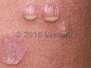
SPD usually presents as crops of small, flaccid pustulovesicles that coalesce into annular or serpiginous configurations on previously normal skin. The lesions most commonly occur symmetrically on the flexural extremities, as well as the axillae, groin, abdomen, and inframammary skin. The pustules dry up after a few days, leaving scaling and crusts that may resemble impetigo. This is followed by new crops of vesicles or pustules developing at the periphery of older lesions, leaving annular or polycyclic lesions. The lesions resolve without scarring but may leave postinflammatory hyperpigmentation. This cycle may repeat itself a few days or weeks later if the patient is not treated.
The etiology of SPD is unknown. SPD has been described in association with IgA paraproteinemia and IgA myeloma. Associations with pyoderma gangrenosum, ulcerative colitis, Crohn disease, rheumatoid arthritis, and systemic lupus erythematosus have also been reported. Additionally, associations with SAPHO syndrome (synovitis, acne, pustulosis, hyperostosis, osteitis), thyroid dysfunction, endocrine tumors, and polycythemia vera have been reported.
Subcorneal pustular dermatosis
Alerts and Notices
Important News & Links
Synopsis

Codes
ICD10CM:
L13.1 – Subcorneal pustular dermatitis
SNOMEDCT:
25147002 – Subcorneal pustular dermatosis
L13.1 – Subcorneal pustular dermatitis
SNOMEDCT:
25147002 – Subcorneal pustular dermatosis
Look For
Subscription Required
Diagnostic Pearls
Subscription Required
Differential Diagnosis & Pitfalls

To perform a comparison, select diagnoses from the classic differential
Subscription Required
Best Tests
Subscription Required
Management Pearls
Subscription Required
Therapy
Subscription Required
Drug Reaction Data
Subscription Required
References
Subscription Required
Last Reviewed:02/05/2020
Last Updated:03/31/2024
Last Updated:03/31/2024
Subcorneal pustular dermatosis