Contents

SynopsisCodesLook ForDiagnostic PearlsDifferential Diagnosis & PitfallsBest TestsManagement PearlsTherapyDrug Reaction DataReferences

View all Images (11)

Hepatocellular carcinoma
Other Resources UpToDate PubMed

Hepatocellular carcinoma

Contributors: Beatriz Malibiran, Jennifer J. Findeis-Hosey MD
Other Resources UpToDate PubMed

Synopsis

Hepatocellular carcinoma (HCC) is the most common primary malignancy of the liver in adults over the age of 65 years worldwide and is considered the third leading cause of cancer death. In the United States, the ratio of incidence of HCC between men and women is 2.5 to 1. The incidence and sex ratio of HCC varies regionally, and the majority of cases are diagnosed in developing nations. The incidence of HCC is highest in East Asia, Southeast Asia, and sub-Saharan Africa where disease is most commonly associated with chronic viral hepatitis B or C infection. Major risk factors for developing HCC include cirrhosis, chronic viral hepatitis, dietary aflatoxin, diabetes, nonalcoholic fatty liver disease (NAFLD), and alcohol abuse. Children are rarely affected; the only known risk factor in this population is chronic hepatitis B carrier status. In the United States, data suggests that the incidence of HCC may have remained stable since 2013, which may be attributed to improvements in antiviral therapy.

Patients are typically asymptomatic in the early stages of the disease. In the United States, right upper quadrant abdominal pain, weight loss, and malaise are common presenting symptoms. Jaundice, ascites, and hepatosplenomegaly may occur in patients with bile duct obstruction. Late findings include portal hypertension, abdominal bleeding, encephalopathy, and liver failure.

The Barcelona Clinic Liver Cancer algorithm is widely used for HCC staging:
  • Very early stage (0): Solitary nodules ≤ 2 cm. Treatment includes ablation or resection.
  • Early stage (A): Solitary nodule > 2 cm OR 2-3 nodules all ≤ 3 cm. Treatment options include resection, transplantation, or ablation.
  • Intermediate stage (B): > 3 nodules OR ≥ 2 nodules if any are > 3 cm. Treat with chemo-embolization.
  • Advanced stage (C): Macrovascular invasion OR extrahepatic spread. Treat with systemic therapies.
  • Terminal stage (D): End-stage liver function. Nontransplantable HCC. Supportive care.

Codes

ICD10CM:
C22.0 – Liver cell carcinoma

SNOMEDCT:
25370001 – Hepatocellular carcinoma

Look For

Subscription Required

Diagnostic Pearls

Subscription Required

Differential Diagnosis & Pitfalls

  • Hepatic adenoma – A benign lesion, generally uncommon but more common among women than men. Risk factors include long-term oral contraceptive use and estrogen exposure during pregnancy. Up to 30% of cases are complicated by bleeding.
  • Dysplastic nodules from cirrhosis
  • Fibrous nodular hyperplasia
  • Cholangiocarcinoma
  • Pyogenic liver abscess
  • Metastatic cancer (colon / rectal, pancreatic, gastric) – distinguish with radiology and biopsy histology

Best Tests

Subscription Required

Management Pearls

Subscription Required

Therapy

Subscription Required

Drug Reaction Data

Below is a list of drugs with literature evidence indicating an adverse association with this diagnosis. The list is continually updated through ongoing research and new medication approvals. Click on Citations to sort by number of citations or click on Medication to sort the medications alphabetically.

Subscription Required

References

Subscription Required

Last Reviewed:04/26/2018
Last Updated:01/31/2023
Copyright © 2023 VisualDx®. All rights reserved.
Hepatocellular carcinoma
A medical illustration showing key findings of Hepatocellular carcinoma : Jaundice, Ascites, Hypoalbuminemia, Hyperbilirubinemia, Splenomegaly, PT prolonged, PLT decreased
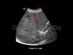
Imaging Studies image of Hepatocellular carcinoma - imageId=8358911. Click to open in gallery.  caption: '<span>Grayscale ultrasound image of the liver demonstrating a heterogeneously hyperechoic mass in the right lobe of the liver. Pathology proven hepatocellular carcinoma.</span>'
Grayscale ultrasound image of the liver demonstrating a heterogeneously hyperechoic mass in the right lobe of the liver. Pathology proven hepatocellular carcinoma.
Copyright © 2023 VisualDx®. All rights reserved.