Onychoschizia (also known as onychoschisis, lamellar dystrophy, and peeling of nails) is characterized by horizontal splits within the nail plate due to loss of intracellular adhesion. Onychoschizia is often seen together with onychorrhexis – longitudinal splitting or ridging of the nail plate – and the 2 findings constitute brittle nail syndrome. Brittle nails affect nearly 20% of the population. Onychoschizia is seen more frequently in women and in the elderly. Frequent wetting and drying of the hands is the most common cause of the loss of intracellular adhesion, and onychoschizia is common among house cleaners, nurses, and hairdressers. Onychoschizia has also been attributed to nail cosmetics (hardeners, polish, polish removers / solvents), nail procedures, and occupational exposure to various chemicals (alkalis, acids, cement, solvents, thioglycolates, salt, sugar solutions). Trauma may also play a role in the pathogenesis of brittle nails.
Onychoschizia has been observed in around 9% of pregnant individuals and represents the second most common nail finding in pregnancy along with onychocryptosis (9%) and leukonychia (24.4%).
Brittle nails have also been associated with systemic diseases, including endocrinopathies, tuberculosis, Sjögren syndrome, and malnutrition. Onychoschizia is a common finding in Sézary syndrome and, along with anonychia and distal notching, may differentiate from the nail findings in early mycosis fungoides. (The most common nail findings seen in early mycosis fungoides are longitudinal ridging, nail thickening, nail fragility, and leukonychia.)
The horizontal splits are most commonly seen in the distal portion of the nail and nail edge; proximal onychoschizia has been observed in patients with psoriasis or lichen planus and in those on oral retinoid therapy.
Toenail onychoschizia has been observed in around one-third of newborns younger than 5 days, and fingernail and toenail involvement is seen in 2.4% of children aged 0-2 years. In the fingernails, thumb sucking is thought to be contributory. It is thought that this splitting is secondary to the thin nails in newborns and children that are easily separated with minor trauma. It resolves as the child gets older.
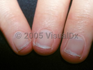
Onychoschizia - Nail and Distal Digit
Alerts and Notices
Important News & Links
Synopsis

Codes
ICD10CM:
L60.3 – Nail dystrophy
SNOMEDCT:
82144009 – Lamellar nail splitting
L60.3 – Nail dystrophy
SNOMEDCT:
82144009 – Lamellar nail splitting
Look For
Subscription Required
Diagnostic Pearls
Subscription Required
Differential Diagnosis & Pitfalls

To perform a comparison, select diagnoses from the classic differential
Subscription Required
Best Tests
Subscription Required
Management Pearls
Subscription Required
Therapy
Subscription Required
Drug Reaction Data
Subscription Required
References
Subscription Required
Last Reviewed:05/01/2019
Last Updated:08/09/2021
Last Updated:08/09/2021
 Patient Information for Onychoschizia - Nail and Distal Digit
Premium Feature
VisualDx Patient Handouts
Available in the Elite package
- Improve treatment compliance
- Reduce after-hours questions
- Increase patient engagement and satisfaction
- Written in clear, easy-to-understand language. No confusing jargon.
- Available in English and Spanish
- Print out or email directly to your patient
Upgrade Today