Contents

SynopsisCodesLook ForDiagnostic PearlsDifferential Diagnosis & PitfallsBest TestsManagement PearlsTherapyDrug Reaction DataReferences

Information for Patients

View all Images (25)

Erythema nodosum in Child
Other Resources UpToDate PubMed

Erythema nodosum in Child

Contributors: Andrina Mamo BS, Andrew Walls MD, Craig N. Burkhart MD, Dean Morrell MD, Susan Burgin MD
Other Resources UpToDate PubMed

Synopsis

Erythema nodosum (EN) is the most common type of septal panniculitis (inflammation of the subcutaneous fat) and results from a delayed hypersensitivity to various stimuli, such as bacterial and viral infections (most commonly Streptococcus and Epstein-Barr virus [EBV], respectively), medications, malignancies, inflammatory bowel disease (see Crohn disease, ulcerative colitis), fungal infections, and collagen vascular diseases. However, in 33%-50% of cases, no associations are found. A detailed list of etiologies is below.

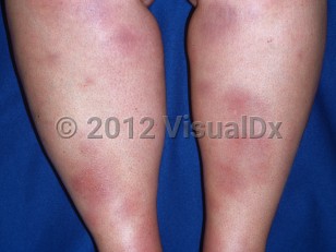
The eruption persists for 3-6 weeks and spontaneously regresses without ulceration, scarring, or atrophy.

EN occurs in boys and girls in equal proportions before puberty (it rarely occurs before age 2); however, after puberty, females are more frequently affected, as in the adult population. Recurrences with reappearance of the precipitating factor(s) are reported.

More commonly associated infections:
Less common infectious associations:
Inflammatory etiologies:
Malignancy (especially leukemia)

Drugs:
  • Oral contraceptives, sulfonamides, penicillins, cephalosporins, macrolide antibiotics
Pregnancy

Codes

ICD10CM:
L52 – Erythema nodosum

SNOMEDCT:
32861005 – Erythema nodosum

Look For

Subscription Required

Diagnostic Pearls

Subscription Required

Differential Diagnosis & Pitfalls

Other forms of panniculitis:
  • Primary infectious panniculitis (see panniculitis) occurs through septic seeding or direct inoculation of a variety of infectious organisms. Tissue culture and polymerase chain reaction (PCR) aids in diagnosis.
  • Subacute migratory panniculitis is often more focal, painless, and unilateral.
  • Nodular vasculitis / erythema induratum is typically on the posterior calves and associated with pulmonary TB.
  • Pancreatic panniculitis favors the lower legs but is often ulcerated.
  • Lupus panniculitis favors the face and upper trunk. Most cases are not associated with systemic lupus erythematosus.
  • Panniculitis of dermatomyositis (abdomen, thighs, arms) is rare but may be the first manifestation of juvenile dermatomyositis.
  • Granulomatous panniculitis is a manifestation of subcutaneous granuloma annulare or sarcoidosis.
  • Poststeroidal panniculitis is rare but exclusive to children. Occurs 1-10 days following withdrawal of systemic corticosteroids. May ulcerate and scar.
  • Cold panniculitis typically presents on the cheeks ("Popsicle panniculitis") but may occur on any body site exposed to cold temperatures. Lateral thigh involvement of horseback riders has been described.
  • Injections, including iatrogenic and factitial.
  • Alpha-1 antitrypsin deficiency panniculitis is extremely rare in children but has been reported as an initial manifestation of the disease. Lesions frequently ulcerate.
  • Lipoatrophic panniculitis of childhood
Also consider:

Best Tests

Subscription Required

Management Pearls

Subscription Required

Therapy

Subscription Required

Drug Reaction Data

Below is a list of drugs with literature evidence indicating an adverse association with this diagnosis. The list is continually updated through ongoing research and new medication approvals. Click on Citations to sort by number of citations or click on Medication to sort the medications alphabetically.

Subscription Required

References

Subscription Required

Last Reviewed:06/26/2022
Last Updated:07/02/2022
Copyright © 2023 VisualDx®. All rights reserved.
Patient Information for Erythema nodosum in Child
Contributors: Medical staff writer

Overview

Erythema nodosum is a skin disorder that produces red bumps under the skin that can be tender. They usually occur due to some underlying disease or use of a medication. The bumps of erythema nodosum can occur anywhere on the body but are most common on the shins. They look like raised bumps or bruises that change from pink to blue-brown.

Who’s At Risk

Erythema nodosum can occur in patients of any age, sex, or race. The inflammation is most common in females, particularly in the age range 18-34 (teens and young adults). In addition, erythema nodosum tends to occur in families who are exposed to the same trigger. While their cause to a particular individual may be unknown, people who receive immunity-weakening drugs (eg, chemotherapy drugs), oral contraceptives, and antibiotics are more likely to develop them. Also, infections and cancers can predispose a person to developing erythema nodosum.

Signs & Symptoms

The most common locations for erythema nodosum include:
  • Shins
  • Knees, ankles, or thighs
  • Forearms
  • Face and neck
Erythema nodosum appears as one or more reddish, warm, and painful lumps (nodules), ranging in size from 1-10 cm.

The onset of erythema nodosum may be associated with fever, generalized achiness, leg swelling, or joint pain.

Individual nodules of erythema nodosum usually last from 1-2 weeks, but new lesions may continue to appear for up to 6 weeks. When an individual lesion of erythema nodosum has resolved, it may leave behind a temporary bruise, which subsequently fades to normal-appearing skin.

Self-Care Guidelines

Although erythema nodosum may occur on its own, it is more often associated with a medication or with an underlying infection or medical condition. Therefore, it is important to see a physician in order to investigate any possible health problems. In the meantime, however, the tenderness of the skin lesions may be alleviated by the following:
  • Restriction of physical activity or bed rest
  • Elevation of the legs (if they are affected)
  • Cool or warm compresses
Generally, people with erythema nodosum do quite well, especially once any underlying medical conditions have been treated.

When to Seek Medical Care

Since erythema nodosum can be associated with underlying infections or health problems, a physician should be consulted within a few days of noticing the skin lesions.

Treatments

After diagnosing you with erythema nodosum, your physician will attempt to identify a possible cause, such as medication, infection, or medical condition. The doctor may order diagnostic tests such as blood work, a chest X-ray, or a throat culture. If an underlying cause is identified, then the physician will treat it appropriately (for example, by discontinuing a medication, prescribing antibiotics for an infection, or treating a health problem).

Once those investigations and treatments are under way, your health care provider may try the following measures to make you more comfortable:
  • Restriction of physical activity or bed rest
  • Anti-inflammatory medications such as ibuprofen or aspirin
  • Potassium iodide
  • Steroids (either taken in pill form or injected directly into the lesions)

References


Bolognia, Jean L., ed. Dermatology, pp.724, 1455, 1552-1555. New York: Mosby, 2003.

Freedberg, Irwin M., ed. Fitzpatrick's Dermatology in General Medicine. 6th ed, pp.1056, 1059, 1849, 1934, 1944. New York: McGraw-Hill, 2003.
Copyright © 2023 VisualDx®. All rights reserved.
Erythema nodosum in Child
A medical illustration showing key findings of Erythema nodosum (Adult) : Arthralgia, Smooth nodules, Smooth plaques
Clinical image of Erythema nodosum - imageId=2299356. Click to open in gallery.  caption: 'Many erythematous and ecchymotic plaques of varying sizes on the lower legs.'
Many erythematous and ecchymotic plaques of varying sizes on the lower legs.
Copyright © 2023 VisualDx®. All rights reserved.